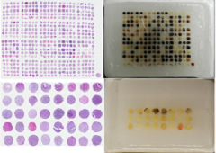
Tissue Microarray
Viewing high magnification power images (100x - 400x) from each tissue array core is now available for almost of all tissue arrays. Click Core Images Tab in the specification sheet page to view the scanned images up to 400x amplification through FullScanViewer provided by TissueArray.com. For example, 616 core high-density array or 208 core high-density array and 96 core array.
Cancer tissue arrays with follow-up data are available. They are kidney, colon, breast, lung and ovarian cancer arrays
Series of cancer survey tissue arrays with TNM, clinical stage and pathology grade, up to 2600 cases of breast cancer were built and listed here.
Test tissue arrays with two identical 12 core arrays on one slide for testing antibody dilutions and experiment conditions in each cancer type are available and listed here.
Disease spectrum tissue arrays (cancer progression) are available. Please browse our Tissue Microarray page to find out from each cancer type only produced by TissueArray.com!
Paraffin Tissue Sections
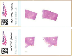
We have pre-cut histology paraffin (FFPE) tissue sections, including normal human paraffin sections HuFPT, human cancer paraffin tissue sections, HuCat and normal mouse/rat paraffin tissue sections.
Formalin fixed, paraffin embedded histology tissue sections are ideal choices for rapidly localizing DNA, RNA and protein markers.
The tissues were fixed by formalin no longer than 48 hours, and then processed to be ready for sectioning.
Two serial tissue sections with 5 µm thickness are mounted on a SuperFrost Plus glass slide.
TissueArray.com histology paraffin tissue section is perfect fo fast detection of genes and proteins expression in specific tissues of different species.


